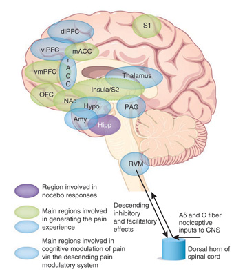
Functional neuroimaging methods have been used to study the neuronal basis of pain and pain perception, demonstrating that painful stimulation activates extensive brain regions initially referred to as the “Pain Matrix.” Although the specificity of pain matrix activation to pain has been questioned, current research studies have demonstrated very high sensitivity and specificity of functional magnetic resonance imaging (fMRI) based regional activation related to pain perception. In addition, positron emission tomography (PET) studies in acute pain have consistently shown increases in cerebral blood flow (CBF) in many of the same brain regions.

* Melzack R (Dec 2001). “Pain and the Neuromatrix in the Brain”.J Dent Educ 65 (12): 1378–82. PMID 11780656
Pain studies using electrophysiological recording (EEG, Evoked Potentials (EP) and MEG) demonstrate shifts in the frequency spectrum in the presence of chronic or induced pain, to the theta and beta frequency bands. Source localization of scalp-recorded EEG data Low-Resolution Electromagnetic Tomographic Analysis (LORETA) in patients with intractable unilateral or bilateral neuropathic pain, when compared to pain-free controls identified similar neural activity sources as reported in the literature with conventional neuroimaging methods.
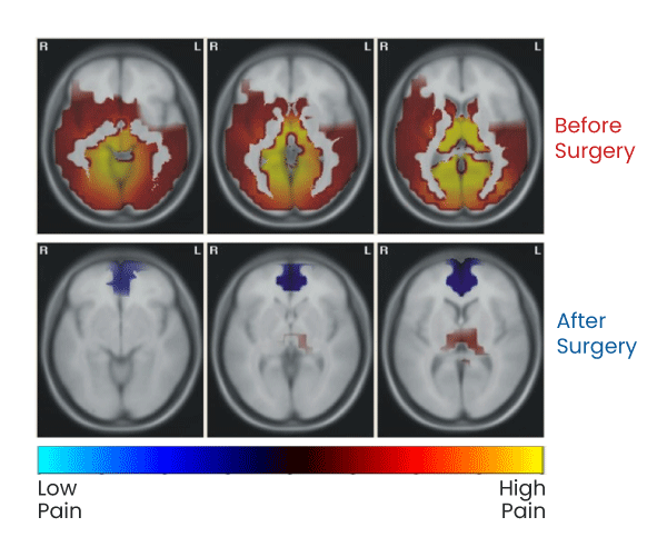
Preliminary data collected at the Brain Research Laboratories, Department of Psychiatry at NYUSOM (under a research contract from PainQx) has been published demonstrating feasibility/proof of concept for an approach which uses QEEG to identify and quantify significant over and under activation of cortical and subcortical regions of interest (ROI) of the brain in a chronic pain population. Using QEEG source localization methods, this published pilot study demonstrated that structures which are thought to be associated with the perception of pain show increased signal in specific frequency bands corresponding to cellular activation when in the painful state, and that evidence of activation diminishes in conjunction with a patient’s reported amelioration of pain.
EEG Based Visualization of High Versus Low Pain States
Overview:
A normal brain looks different than a brain in pain as seen in the following EEG Source Localization images
Patient Case Study:
Patient with chronic neuropathic pain caused by nerve compression in the spine.
Top Panel: Patient Before Surgical Decompression
Highly activated brain regions are shown. Patient reported pain score was 4 [out of 10]
Bottom Panel: Patient After Surgical Decompression
Minimally activated brain regions are shown. After surgery, patient reported pain score was 0 [out of 10]


